The Cause Of Bunions?
Overview
 A bunion is an abnormal bump, or bony enlargement that forms at the base of your big toe. In the early stages, the bunion may be small and the side of your foot may be slightly swollen. As it progresses, the bump gets larger and becomes increasingly painful. Your big toe may point towards your other toes, rather than straight. Shoes may be uncomfortable to wear and add to the irritation of the joint.
Causes
Bunions are a common problem that can cause foot pain and difficulty wearing shoes. Bunions occur in about 30% of the population of most Western countries. They are seen most commonly in women and become more common as people get older. Patients with bunions generally have one of two problems that can cause pain. As the big toe becomes more and more angled (pointing toward the other toes), the base of the toe becomes more and more prominent, forming the bunion. The bunion forms in part because of the new angle of the toe, and in part due to inflammation over the bunion surface. As the inflammation worsens, people can experience pain with shoe wear and walking. The big toe may eventually come to lie over, or more commonly under, the second toe. This may cause further irritation while wearing shoes and more pain. The second toe of patients who have bunions commonly forms a hammer toe.
Symptoms
Bunions may or may not cause symptoms. A frequent symptom is foot pain in the involved area when walking or wearing shoes; rest relieves this pain. A bunion causes enlargement of the base of the big toe and is usually associated with positioning of the big toe toward the smaller toes. Shoe pressure in this area can cause interment pain while the development of arthritis in more severe bunions can lead to chronic pain. Bunions that cause marked pain are often associated with swelling of the soft tissues, redness, and local tenderness. It is important to note that, in postpubertal men and postmenopausal women, pain at the base of the big toe can be caused by gout and gouty arthritis that is similar to the pain caused by bunions.
Diagnosis
Although bunions are usually obvious from the pain and unusual shape of the toe, further investigation is often advisable. Your doctor will usually send you for X-rays to determine the extent of the deformity. Blood tests may be advised to see if some type of arthritis could be causing the pain. Based on this evaluation, your doctor can determine whether you need orthopaedic shoes, medication, surgery or other treatment.
Non Surgical Treatment
Wide toe box, bunion pads, orthotics, or a combination. Mild discomfort may lessen by wearing a shoe with a wide toe box or with stretchable material. If not, bunion pads purchased in most pharmacies can shield the painful area. Orthotics can also be prescribed to redistribute and relieve pressure from the affected articulation. If conservative therapy fails, surgery aimed at correcting abnormal bony alignments and restoring joint mobility should be considered. If the patient is unwilling to wear large, wider shoes to accommodate the bunion because they are unattractive, surgery can be considered; however, patients should be told that orthotic devices should be worn after surgery to reduce the risk of recurrence. For bursitis, bursal aspiration and injection of a corticosteroid are indicated. For osteoarthritic synovitis, oral NSAIDs or an intra-articular injection of a corticosteroid/anesthetic solution reduces symptoms. For hallux limitus or hallux rigidus, treatment aims to preserve joint mobility by using passive stretching exercises, which occasionally require injection of a local anesthetic to relieve muscle spasm. Sometimes surgical release of contractures is necessary. 
Surgical Treatment
Bunion surgery is occasionally required when the bunion deformity is too advanced for conservative treatment to work. Your surgeon will usually cut an angular section from the bone to correct the alignment. In some cases, multiple toes may need to be straighten.
What Is Over-Pronation Of The Feet
Overview
Overpronation is the exaggerated inward rolling of the foot and ankle, which can lead to a collapsed arch and flat feet. Many people overpronate and do not even realize it; one way to tell is to simply look at the foot and see how it is placed on the ground when standing in a neutral position. Another way is to wet the bottom of the foot and step on a piece of paper. If the entire imprint of the foot is shown, it means you overpronate.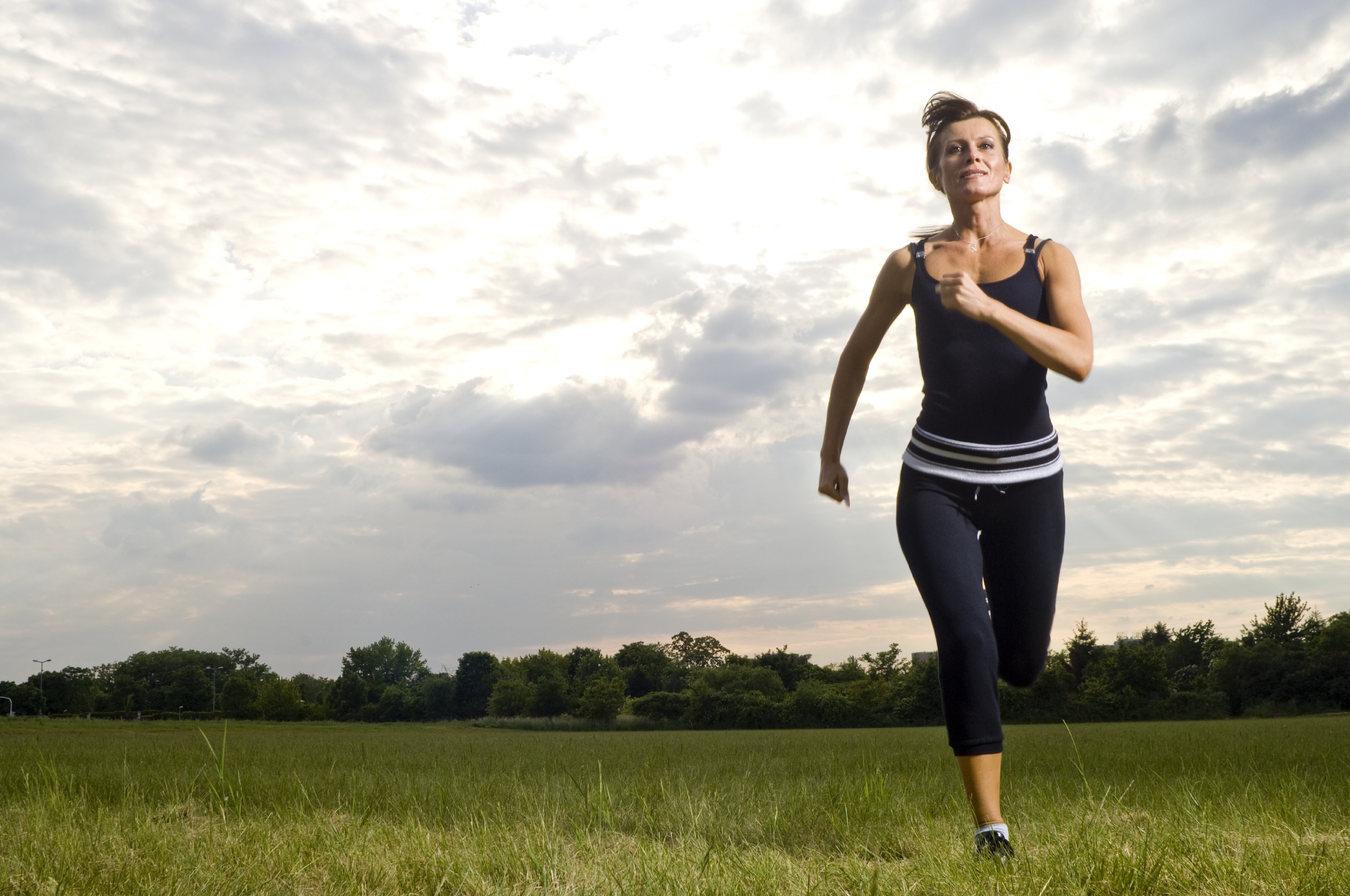
Causes
Flat feet don't automatically mean you have a problem. The problem can be divided into a flexible flat foot or rigid flat foot. The rigid flat foot is one that does not change shape when the foot becomes weight bearing. i.e. it does not go through the excessive motion of pronation. Generally speaking this foot does not provide too many problems. The flexible flat foot is the type that when it becomes weight bearing the foot and ankle tends to roll in (pronates) too far. This type of person will often say I have great arches but when I stand up much of this arch disappears as the foot excessively pronates When the foot is excessively pronating and causing problems like sore ankles, feet or knees when standing or exercising then arch support is extremely important to restore the foot structure.
Symptoms
Overpronation causes alterations in proper muscle recruitment patterns leading to tightness in the outside of the ankle (lateral gastrocnemius, soleus, and peroneals). This tightness can lead to weakness in the opposing muscles such as the medial gastrocnemius, anterior tibialis, and posterior tibialis. If these muscles are weak, they will not be able to keep the knee in proper alignment, causing the valgus position. All this tightness and weakness can cause pain within the ankle, calf, and knee region. And it can send imbalance and pain all the way up to the upper back, if deep core strength is lacking and can't hold the pelvis in neutral.
Diagnosis
When sitting, an over-pronating foot appears quite normal, i.e. showing a normal arch with room under the underside of the foot. The moment you get up and put weight on your feet the situation changes: the arches lower and the ankle slightly turns inwards. When you walk or run more weight is placed on the feet compared to standing and over-pronation will become more evident. When walking barefoot on tiles or timber floors over-pronation is more visible, compared to walking on carpet or grass.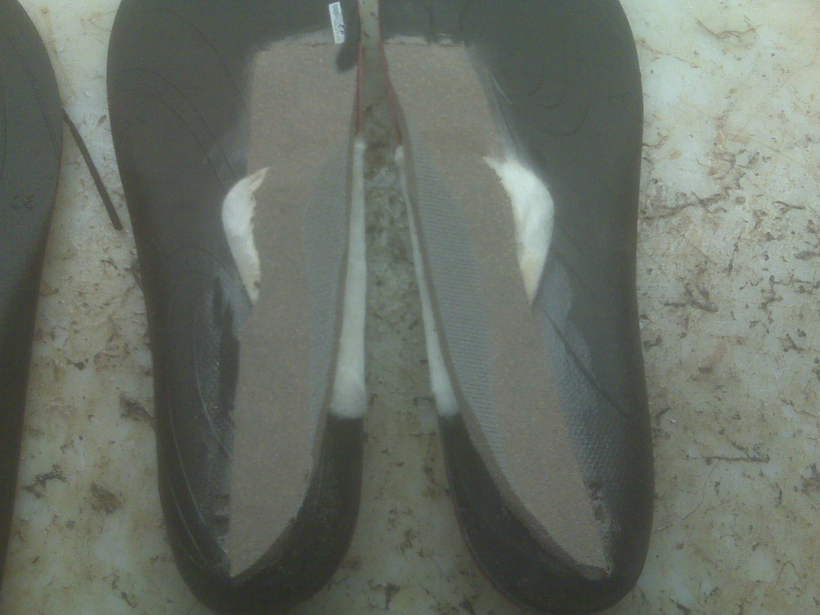
Non Surgical Treatment
Mild cases of Overpronation may be controlled or corrected with a supportive shoe that offers medial support to the foot along with a strong heel counter to control excessive motion at the heel starting with heel strike. In mild cases with no abnormal mechanical pressures, an over the counter orthotic with heel cup and longitudinal or medial arch support to keep the foot from progressing past neutral may help to realign the foot. A Custom foot orthotic with heel cup and longitudinal arch support to help correct position of the foot as it moves through motion. Heel wedges may also assist in correcting motion.
Prevention
Custom-made orthotics supports not only the arch as a whole, but also each individual bone and joint that forms the arch. It is not enough to use an over-the-counter arch support, as these generic devices will not provide the proper support to each specific structure of the arch and foot. Each pronated foot?s arch collapses differently and to different degrees. The only way to provide the support that you may need is with a custom-made device. This action of the custom-made orthotic will help to prevent heel spurs, plantar fasciitis, calluses, arch pain, and weakness of the entire foot.
Physiotherapy For Calcaneal Apophysitis
Overview
Sever's disease occurs in children when the growth plate (which is the growing part of the heel) is injured. The foot is one of the first body parts to grow to full size. This usually occurs in early puberty. During this time, bones often grow faster than muscles and tendons. As a result, muscles and tendons become tight. The heel area is less flexible. During weight-bearing activity (activity performed while standing), the tight heel tendons may put too much pressure at the back of the heel (where the Achilles tendon attaches). This can injure the heel and cause Sever's disease.
Causes
The usual cause is directly related to overuse of the bone and tendons in the heel. This can come from playing sports or anything that involves a lot of heel movement. It can be associated with starting a new sport, or the start of a new season, or too much weight bearing on the heel. Also, excessive traction could cause this, since the bones and tendons are still developing. Many children who over pronate their feet exhibit symptoms and in most patients, it usually involves both heels.
Symptoms
Sharp pain will be present in the affected heel (or both heels), especially while running or walking. Pain can be heightened following activity. The area will be tender to the touch and usually becomes inflamed or reddened. It may also be painful to press on the heel with a finger from the back or to squeeze the sides together; the latter is particularly common. You might notice stiffness in some of the surrounding muscles, making regular movements more difficult to achieve. This and the pain can manifest physically in abnormal practices like tiptoeing or limping. In some cases a lump can be detected on the back of the heel, though it may be so small as to defy detection.
Diagnosis
Sever's disease is diagnosed based on a doctor?s physical examination of the lower leg, ankle, and foot. If the diagnosis is in question, the doctor may order X-rays or an MRI to determine if there are other injuries that may be causing the heel pain.
Non Surgical Treatment
Stretching programs. Strengthening exercises. Exercise and training modification. Orthotic therapy. In rare cases, where fragmentation of the apophysis exists and pain fails to subside with traditional treatments then immobilization of the foot and ankle with a short leg pneumatic walker(walking cast) is indicated.
Which Are The Principal Causes Of Posterior Tibial Tendon Dysfunction (PTTD) ?
Overview
Adult acquired flatfoot deformity (AAFD) is a painful condition resulting from the collapse of the longitudinal (lengthwise) arch of the foot. As the name suggests, this condition is not present at birth or during childhood. It occurs after the skeleton is fully matured. In the past it was referred to a posterior tibial tendon dysfunction (or insufficiency). But the name was changed because the condition really describes a wide range of flatfoot deformities. AAFD is most often seen in women between the ages of 40 and 60. This guide will help you understand how the problem develops, how doctors diagnose the condition, what treatment options are available. 
Causes
Overuse of the posterior tibial tendon is often the cause of PTTD. In fact, the symptoms usually occur after activities that involve the tendon, such as running, walking, hiking, or climbing stairs.
Symptoms
Often, this condition is only present in one foot, but it can affect both. Adult acquired flatfoot symptoms vary, but can swelling of the foot's inner side and aching heel and arch pain. Some patients experience no pain, but others may experience severe pain. Symptoms may increase during long periods of standing, resulting in fatigue. Symptoms may change over time as the condition worsens. The pain may move to the foot's outer side, and some patients may develop arthritis in the ankle and foot.
Diagnosis
Examination by your foot and ankle specialist can confirm the diagnosis for most patients. An ultrasound exam performed in the office setting can evaluate the status of the posterior tibial tendon, the tendon which is primarily responsible for supporting the arch structure of the foot.
Non surgical Treatment
Initial treatment consists of supporting the medial longitudinal arch (running the length of the foot) to relieve strain on the medial soft tissues. The most effective way to relieve pain on the tendon is to use a boot or brace, and once tenderness and pain has resolved, an orthotic device. A boot, brace, or orthotic has not been shown to correct or even prevent the progression of deformity. Orthotics can alleviate symptoms and may slow the progression of deformity, particularly if mild. The deformity may progress despite orthotics. 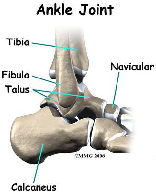
Surgical Treatment
If conservative treatments don?t work, your doctor may recommend surgery. Several procedures can be used to treat posterior tibial tendon dysfunction; often more than one procedure is performed at the same time. Your doctor will recommend a specific course of treatment based on your individual case. Surgical options include. Tenosynovectomy. In this procedure, the surgeon will clean away (debride) and remove (excise) any inflamed tissue surrounding the tendon. Osteotomy. This procedure changes the alignment of the heel bone (calcaneus). The surgeon may sometimes have to remove a portion of the bone. Tendon transfer: This procedure uses some fibers from another tendon (the flexor digitorum longus, which helps bend the toes) to repair the damaged posterior tibial tendon. Lateral column lengthening, In this procedure, the surgeon places a small wedge-shaped piece of bone into the outside of the calcaneus. This helps realign the bones and recreates the arch. Arthrodesis. This procedure welds (fuses) one or more bones together, eliminating movement in the joint. This stabilizes the hindfoot and prevents the condition from progressing further.
Foot Pain In The Arch And Ankle
Overview
The arch of the foot is made from a complex series of bones tendons and ligaments. According to podiatric practice there is only one important arch of the foot on the medial side. The height of the arch varies and having a flat foot or high arched foot is inherited. The arch acts to dissipate forces and store energy within the foot. Most young children have flat feet and this is because the arch has fatty tissue overlying it. Normally an arch appears after the age of 5-6 years. Even if you remain flat footed this might be normal for you and your family. Just because the arch is low does not mean that any treatment is required. Many world class athletes have flat feet. If the arch is painful or the arch of one foot suddenly drops then investigation and treatment may be required. The arch might drop acutely due to a tear in a tendon or the movement of a bone within the arch. A scan and X Ray may be indicated and treatment ranging from insoles to surgery may be required. If you have a high arched foot this is likely to have been inherited. In extreme form this can cause pain in the ball of the foot and is often associated with a tight Achilles tendon and a tendency to walk on the outside of your foot. In most people there are no symptoms and in others there may pain along the arch and pain on the outside of your ankle. Both these foot types are often mild and require no treatment, but if symptoms occur a full bio-mechanical examination will often reveal a mechanical cause to your pain. Often the treatment may be simple stretches and orthoses.

Causes
Over-stretching or tearing the arch pad causes the dull ache that is associated with arch pain. Pain is most keenly felt when weight is put onto the foot, or when pushing off the foot into the next stride. The pain of foot arch strain can be particularly acute after sitting for a long time, or when getting up in the morning. Those most susceptible to the condition are people with very high, rigid arches. The injury is also common in middle-aged people who have been inactive for a long period of time before suddenly increasing their physical activity. The injury is particularly common in runners and joggers.
Symptoms
Flat feet don't usually cause problems, but they can put a strain on your muscles and ligaments (ligaments link two bones together at a joint). This may cause pain in your legs when you walk. If you have flat feet, you may experience pain in any of the following areas, the inside of your ankle, the arch of your foot, the outer side of your foot, the calf, the knee, hip or back, Some people with flat feet find that their weight is distributed unevenly, particularly if their foot rolls inwards too much (overpronates). If your foot overpronates, your shoes are likely to wear out quickly. Overpronation can also damage your ankle joint and Achilles tendon (the large tendon at the back of your ankle). See your GP if you or your child has flat feet and your feet are painful, even when wearing supportive, well-fitting shoes, shoes wear out very quickly, feet appear to be getting flatter, feet are weak, numb or stiff, Your GP may refer you to a podiatrist (foot specialist).
Diagnosis
Your doctor may order imaging tests to help make sure your heel pain is caused by plantar fasciitis and not another problem. X-rays provide clear images of bones. They are useful in ruling out other causes of heel pain, such as fractures or arthritis. Heel spurs can be seen on an x-ray. Other imaging tests, such as magnetic resonance imaging (MRI) and ultrasound, are not routinely used to diagnose plantar fasciitis. They are rarely ordered. An MRI scan may be used if the heel pain is not relieved by initial treatment methods.
Non Surgical Treatment
Once the severity and cause of arch and foot pain is determined, a course of corrective and rehabilitative actions can be started. Therapists may use machines and manual therapies to reduce pain and increase circulation to the area to promote healing. Maintenance of fitness levels via modification of activity may be prescribed. Substitute activities that may aggravate the pain and soreness with other activities; for instance, running causes the body to have multiple impacts with the ground, but the use of bicycling, elliptical trainers, step machines, swimming, or ski machines eliminates impact and allows you to continue to maintain and improve your fitness levels. Take medications to help reduce pain and inflammation. Follow up with your doctor until you are better.

Surgical Treatment
Surgery for flat feet is separated into three kinds: soft tissue procedures, bone cuts, and bone fusions. Depending on the severity of the flat foot, a person?s age, and whether or not the foot is stiff determines just how the foot can be fixed. In most cases a combination of procedures are performed. With flexible flat feet, surgery is geared at maintaining the motion of the foot and recreating the arch. Commonly this may involve tendon repairs along the inside of the foot to reinforce the main tendon that lifts the arch. When the bone collapse is significant, bone procedures are included to physically rebuild the arch, and realign the heel. The presence of bunions with flat feet is often contributing to the collapse and in most situations requires correction. With rigid flat feet, surgery is focused on restoring the shape of the foot through procedures that eliminate motion. In this case, motion does not exist pre-operatively, so realigning the foot is of utmost importance. The exception, are rigid flat feet due to tarsal coalition (fused segment of bone) in the back of the foot where freeing the blockage can restore function.
Prevention
Early in the treatment of arch pain, consideration needs to be given to the cause and strategies put in place to prevent it happening again. Advice should be sought on the adequacy of footwear. Stretching exercises should be continued long after the symptoms are gone. Foot orthoses should be used if structural imbalances are present. Activity levels and types of activities (occupational and sporting) need to be considered and modified accordingly.
Stretching Exercises
Massage therapy is a great way to loosen muscles and help improve mobility in in your feet. As many people with foot pain have discovered, tight muscles in your legs or back can lead to tense foot muscles. All those muscles are connected, so tension in your back can cause tension in your legs which can pull the tendons in your feet and cause stiffness and pain. Getting acupuncture or a professional full body massage are probably the best ways to deal with this, but there are also some simple tricks you can do at home to help keep muscles limber. These are great for loosening up and improving circulation, both before and after exercise. Place a tennis ball under the arch of your bare foot and roll it around, stretching the muscles in your foot and promoting blood flow. You can also roll the ball under your calves and upper legs to work out stiffness and knots. If you feel the tennis ball is too easy, try a lacrosse ball for deeper massaging. This is also demonstrated in the exercise video above. Use a foam roller, those big overpriced rolls of foam that are now available in every department and sporting goods store are fantastic for self-massage (why a roll of foam costs $30 is beyond us, but they do work wonders-our advice is to not waste money on the more expensive fancy grooved ones because even the simplest rollers work great). The exercises you can do with foam rollers seem to be endless, and there are literally hundreds of free videos online showing how to use them to massage every part of your body. Here's one we picked out that specifically targets foot and leg muscles related to arches and plantar fasciitis.
The arch of the foot is made from a complex series of bones tendons and ligaments. According to podiatric practice there is only one important arch of the foot on the medial side. The height of the arch varies and having a flat foot or high arched foot is inherited. The arch acts to dissipate forces and store energy within the foot. Most young children have flat feet and this is because the arch has fatty tissue overlying it. Normally an arch appears after the age of 5-6 years. Even if you remain flat footed this might be normal for you and your family. Just because the arch is low does not mean that any treatment is required. Many world class athletes have flat feet. If the arch is painful or the arch of one foot suddenly drops then investigation and treatment may be required. The arch might drop acutely due to a tear in a tendon or the movement of a bone within the arch. A scan and X Ray may be indicated and treatment ranging from insoles to surgery may be required. If you have a high arched foot this is likely to have been inherited. In extreme form this can cause pain in the ball of the foot and is often associated with a tight Achilles tendon and a tendency to walk on the outside of your foot. In most people there are no symptoms and in others there may pain along the arch and pain on the outside of your ankle. Both these foot types are often mild and require no treatment, but if symptoms occur a full bio-mechanical examination will often reveal a mechanical cause to your pain. Often the treatment may be simple stretches and orthoses.
Causes
Over-stretching or tearing the arch pad causes the dull ache that is associated with arch pain. Pain is most keenly felt when weight is put onto the foot, or when pushing off the foot into the next stride. The pain of foot arch strain can be particularly acute after sitting for a long time, or when getting up in the morning. Those most susceptible to the condition are people with very high, rigid arches. The injury is also common in middle-aged people who have been inactive for a long period of time before suddenly increasing their physical activity. The injury is particularly common in runners and joggers.
Symptoms
Flat feet don't usually cause problems, but they can put a strain on your muscles and ligaments (ligaments link two bones together at a joint). This may cause pain in your legs when you walk. If you have flat feet, you may experience pain in any of the following areas, the inside of your ankle, the arch of your foot, the outer side of your foot, the calf, the knee, hip or back, Some people with flat feet find that their weight is distributed unevenly, particularly if their foot rolls inwards too much (overpronates). If your foot overpronates, your shoes are likely to wear out quickly. Overpronation can also damage your ankle joint and Achilles tendon (the large tendon at the back of your ankle). See your GP if you or your child has flat feet and your feet are painful, even when wearing supportive, well-fitting shoes, shoes wear out very quickly, feet appear to be getting flatter, feet are weak, numb or stiff, Your GP may refer you to a podiatrist (foot specialist).
Diagnosis
Your doctor may order imaging tests to help make sure your heel pain is caused by plantar fasciitis and not another problem. X-rays provide clear images of bones. They are useful in ruling out other causes of heel pain, such as fractures or arthritis. Heel spurs can be seen on an x-ray. Other imaging tests, such as magnetic resonance imaging (MRI) and ultrasound, are not routinely used to diagnose plantar fasciitis. They are rarely ordered. An MRI scan may be used if the heel pain is not relieved by initial treatment methods.
Non Surgical Treatment
Once the severity and cause of arch and foot pain is determined, a course of corrective and rehabilitative actions can be started. Therapists may use machines and manual therapies to reduce pain and increase circulation to the area to promote healing. Maintenance of fitness levels via modification of activity may be prescribed. Substitute activities that may aggravate the pain and soreness with other activities; for instance, running causes the body to have multiple impacts with the ground, but the use of bicycling, elliptical trainers, step machines, swimming, or ski machines eliminates impact and allows you to continue to maintain and improve your fitness levels. Take medications to help reduce pain and inflammation. Follow up with your doctor until you are better.
Surgical Treatment
Surgery for flat feet is separated into three kinds: soft tissue procedures, bone cuts, and bone fusions. Depending on the severity of the flat foot, a person?s age, and whether or not the foot is stiff determines just how the foot can be fixed. In most cases a combination of procedures are performed. With flexible flat feet, surgery is geared at maintaining the motion of the foot and recreating the arch. Commonly this may involve tendon repairs along the inside of the foot to reinforce the main tendon that lifts the arch. When the bone collapse is significant, bone procedures are included to physically rebuild the arch, and realign the heel. The presence of bunions with flat feet is often contributing to the collapse and in most situations requires correction. With rigid flat feet, surgery is focused on restoring the shape of the foot through procedures that eliminate motion. In this case, motion does not exist pre-operatively, so realigning the foot is of utmost importance. The exception, are rigid flat feet due to tarsal coalition (fused segment of bone) in the back of the foot where freeing the blockage can restore function.
Prevention
Early in the treatment of arch pain, consideration needs to be given to the cause and strategies put in place to prevent it happening again. Advice should be sought on the adequacy of footwear. Stretching exercises should be continued long after the symptoms are gone. Foot orthoses should be used if structural imbalances are present. Activity levels and types of activities (occupational and sporting) need to be considered and modified accordingly.
Stretching Exercises
Massage therapy is a great way to loosen muscles and help improve mobility in in your feet. As many people with foot pain have discovered, tight muscles in your legs or back can lead to tense foot muscles. All those muscles are connected, so tension in your back can cause tension in your legs which can pull the tendons in your feet and cause stiffness and pain. Getting acupuncture or a professional full body massage are probably the best ways to deal with this, but there are also some simple tricks you can do at home to help keep muscles limber. These are great for loosening up and improving circulation, both before and after exercise. Place a tennis ball under the arch of your bare foot and roll it around, stretching the muscles in your foot and promoting blood flow. You can also roll the ball under your calves and upper legs to work out stiffness and knots. If you feel the tennis ball is too easy, try a lacrosse ball for deeper massaging. This is also demonstrated in the exercise video above. Use a foam roller, those big overpriced rolls of foam that are now available in every department and sporting goods store are fantastic for self-massage (why a roll of foam costs $30 is beyond us, but they do work wonders-our advice is to not waste money on the more expensive fancy grooved ones because even the simplest rollers work great). The exercises you can do with foam rollers seem to be endless, and there are literally hundreds of free videos online showing how to use them to massage every part of your body. Here's one we picked out that specifically targets foot and leg muscles related to arches and plantar fasciitis.
Dealing With Achilles Tendonitis Suffering
Overview
 Achilles-tendinitis is characterized by inflammation and pain at the Achilles tendon (back of the ankle). This tendon, sometimes called the heel cord, is the tendon attachment of the calf muscles from the leg and knee to the heel. This structure is important in standing on your toes or in the pushing-off phase of walking, running, or jumping.Achilles-tendinitis is usually a grade 1 or 2 strain of the tendon. A grade 1 strain is a mild strain. There is a slight pull of the tendon without obvious tendon tearing. (There is microscopic tendon tearing.) There is no loss of strength, and the tendon is the correct length. A grade 2 strain is a moderate strain. There is tearing of tendon fibers within the substance of the tendon or where the tendon attaches to muscle or bone. The length of the tendon or whole muscle-tendon-bone unit is increased, and there is usually decreased strength. A grade 3 strain is a complete rupture of the tendon.
Causes
Some of the causes of Achilles tendonitis include, overuse injury - this occurs when the Achilles tendon is stressed until it develops small tears. Runners seem to be the most susceptible. People who play sports that involve jumping, such as basketball, are also at increased risk. Arthritis - Achilles tendonitis can be a part of generalised inflammatory arthritis, such as ankylosing spondylitis or psoriatic arthritis. In these conditions, both tendons can be affected. Foot problems - some people with flat feet or hyperpronated feet (feet that turn inward while walking) are prone to Achilles tendonitis. The flattened arch pulls on calf muscles and keeps the Achilles tendon under tight strain. This constant mechanical stress on the heel and tendon can cause inflammation, pain and swelling of the tendon. Being overweight can make the problem worse. Footwear - wearing shoes with minimal support while walking or running can increase the risk, as can wearing high heels. Overweight and obesity - being overweight places more strain on many parts of the body, including the Achilles tendon. Quinolone antibiotics - can in some instances be associated with inflammatory tenosynovitis and, if present, will often be bilateral (both Achilles), coming on soon after exposure to the drug.
Symptoms
Achilles tendonitis typically starts off as a dull stiffness in the tendon, which gradually goes away as the area gets warmed up. It may get worse with faster running, uphill running, or when wearing spikes and other low-heeled running shoes. If you continue to train on it, the tendon will hurt more sharply and more often, eventually impeding your ability even to jog lightly. About two-thirds of Achilles tendonitis cases occur at the ?midpoint? of the tendon, a few inches above the heel. The rest are mostly cases of ?insertional? Achilles tendonitis, which occurs within an inch or so of the heelbone. Insertional Achilles tendonitis tends to be more difficult to get rid of, often because the bursa, a small fluid-filled sac right behind the tendon, can become irritated as well.
Diagnosis
During an examination of the foot and ankle, you doctor will look for the following signs, Achilles tendon swelling or thickening. Bone spurs appearing at the lower part of the tendon at the back of the hell. Pain at the middle or lower area of the Achilles tendon. Limited range of motion of the foot and ankle, and a decreased ability to flex the foot. Your doctor may perform imaging tests, such as X-rays and MRI scans, to make a diagnosis of Achilles tendinitis. X-rays show images of the bones and can help the physician to determine if the Achilles tendon has become hardened, which indicated insertional Achilles tendinitis. MRI scans may not be necessary, but they are important guides if you are recommended to have surgical treatment. An MRI can show the severity of the damage and determine what kind of procedure would be best to address the condition.
Nonsurgical Treatment
There is insufficient evidence from randomised controlled trials to determine which method of treatment is the most appropriate for the treatment of acute or chronic Achilles tendonitis. The patient should abstain from aggravating activities, but with a minimum of rest in order to preserve overall fitness. Possible treatments are non-steroidal anti-inflammatory drugs (NSAIDs), ice, rest, increased warm-up/stretching exercises, physiotherapy and heel lifts (orthotic devices - used on both sides to prevent a gait imbalance). Other treatments evaluated in a Cochrane review were heparin, steroid injections, glycosaminoglycan sulfate, Actovegin?, and topical laser treatment. There was no clear evidence of benefit from these. Casting is an option for resistant Achilles tendonitis. Drugs - analgesics and NSAIDs. Surgery is sometimes used for resistant Achilles tendonitis, but usually as a last resort. Other recently reported treatments include continuing sporting activity in conjunction with rehabilitation, low-energy shock wave therapy[4] and topical glyceryl trinitrate .

Surgical Treatment
Many people don't realize that Achilles tendon surgery can be very traumatic to your body. The type of trauma you experience after surgery can be compared to what you go through when you first injured your Achilles tendon. During the first 24 to 72 hours after the surgery your ankle will be tender, swollen and very painful. Your leg will be weak and unstable making it impossible for you to put weight on your leg without some kind of help. This is why your doctor or surgeon will have you outfitted for a cast, ankle brace and/or crutches before the procedure. When you are relying on a cast/brace and crutches your Achilles tendon is less likely to be as active as it once was. This is usually why atrophy (loss) of your lower leg muscles (specifically your calf muscle) happens. In general, more than 80%* of people who undergo surgery for an injured Achilles Tendon are able to return to their active lifestyle. In order to avoid re-injury, it is important to commit to a regular conservative therapy routine.
Prevention
To prevent Achilles tendonitis or tendonosis from recurring after surgical or non-surgical treatment, the foot and ankle surgeon may recommend strengthening and stretching of the calf muscles through daily exercises. Wearing proper shoes for the foot type and activity is also important in preventing recurrence of the condition.
Achilles-tendinitis is characterized by inflammation and pain at the Achilles tendon (back of the ankle). This tendon, sometimes called the heel cord, is the tendon attachment of the calf muscles from the leg and knee to the heel. This structure is important in standing on your toes or in the pushing-off phase of walking, running, or jumping.Achilles-tendinitis is usually a grade 1 or 2 strain of the tendon. A grade 1 strain is a mild strain. There is a slight pull of the tendon without obvious tendon tearing. (There is microscopic tendon tearing.) There is no loss of strength, and the tendon is the correct length. A grade 2 strain is a moderate strain. There is tearing of tendon fibers within the substance of the tendon or where the tendon attaches to muscle or bone. The length of the tendon or whole muscle-tendon-bone unit is increased, and there is usually decreased strength. A grade 3 strain is a complete rupture of the tendon.
Causes
Some of the causes of Achilles tendonitis include, overuse injury - this occurs when the Achilles tendon is stressed until it develops small tears. Runners seem to be the most susceptible. People who play sports that involve jumping, such as basketball, are also at increased risk. Arthritis - Achilles tendonitis can be a part of generalised inflammatory arthritis, such as ankylosing spondylitis or psoriatic arthritis. In these conditions, both tendons can be affected. Foot problems - some people with flat feet or hyperpronated feet (feet that turn inward while walking) are prone to Achilles tendonitis. The flattened arch pulls on calf muscles and keeps the Achilles tendon under tight strain. This constant mechanical stress on the heel and tendon can cause inflammation, pain and swelling of the tendon. Being overweight can make the problem worse. Footwear - wearing shoes with minimal support while walking or running can increase the risk, as can wearing high heels. Overweight and obesity - being overweight places more strain on many parts of the body, including the Achilles tendon. Quinolone antibiotics - can in some instances be associated with inflammatory tenosynovitis and, if present, will often be bilateral (both Achilles), coming on soon after exposure to the drug.
Symptoms
Achilles tendonitis typically starts off as a dull stiffness in the tendon, which gradually goes away as the area gets warmed up. It may get worse with faster running, uphill running, or when wearing spikes and other low-heeled running shoes. If you continue to train on it, the tendon will hurt more sharply and more often, eventually impeding your ability even to jog lightly. About two-thirds of Achilles tendonitis cases occur at the ?midpoint? of the tendon, a few inches above the heel. The rest are mostly cases of ?insertional? Achilles tendonitis, which occurs within an inch or so of the heelbone. Insertional Achilles tendonitis tends to be more difficult to get rid of, often because the bursa, a small fluid-filled sac right behind the tendon, can become irritated as well.
Diagnosis
During an examination of the foot and ankle, you doctor will look for the following signs, Achilles tendon swelling or thickening. Bone spurs appearing at the lower part of the tendon at the back of the hell. Pain at the middle or lower area of the Achilles tendon. Limited range of motion of the foot and ankle, and a decreased ability to flex the foot. Your doctor may perform imaging tests, such as X-rays and MRI scans, to make a diagnosis of Achilles tendinitis. X-rays show images of the bones and can help the physician to determine if the Achilles tendon has become hardened, which indicated insertional Achilles tendinitis. MRI scans may not be necessary, but they are important guides if you are recommended to have surgical treatment. An MRI can show the severity of the damage and determine what kind of procedure would be best to address the condition.
Nonsurgical Treatment
There is insufficient evidence from randomised controlled trials to determine which method of treatment is the most appropriate for the treatment of acute or chronic Achilles tendonitis. The patient should abstain from aggravating activities, but with a minimum of rest in order to preserve overall fitness. Possible treatments are non-steroidal anti-inflammatory drugs (NSAIDs), ice, rest, increased warm-up/stretching exercises, physiotherapy and heel lifts (orthotic devices - used on both sides to prevent a gait imbalance). Other treatments evaluated in a Cochrane review were heparin, steroid injections, glycosaminoglycan sulfate, Actovegin?, and topical laser treatment. There was no clear evidence of benefit from these. Casting is an option for resistant Achilles tendonitis. Drugs - analgesics and NSAIDs. Surgery is sometimes used for resistant Achilles tendonitis, but usually as a last resort. Other recently reported treatments include continuing sporting activity in conjunction with rehabilitation, low-energy shock wave therapy[4] and topical glyceryl trinitrate .
Surgical Treatment
Many people don't realize that Achilles tendon surgery can be very traumatic to your body. The type of trauma you experience after surgery can be compared to what you go through when you first injured your Achilles tendon. During the first 24 to 72 hours after the surgery your ankle will be tender, swollen and very painful. Your leg will be weak and unstable making it impossible for you to put weight on your leg without some kind of help. This is why your doctor or surgeon will have you outfitted for a cast, ankle brace and/or crutches before the procedure. When you are relying on a cast/brace and crutches your Achilles tendon is less likely to be as active as it once was. This is usually why atrophy (loss) of your lower leg muscles (specifically your calf muscle) happens. In general, more than 80%* of people who undergo surgery for an injured Achilles Tendon are able to return to their active lifestyle. In order to avoid re-injury, it is important to commit to a regular conservative therapy routine.
Prevention
To prevent Achilles tendonitis or tendonosis from recurring after surgical or non-surgical treatment, the foot and ankle surgeon may recommend strengthening and stretching of the calf muscles through daily exercises. Wearing proper shoes for the foot type and activity is also important in preventing recurrence of the condition.
Fallen Arches Causes, Signs Or Symptoms And Therapy
Overview

Flat feet and fallen arches are common conditions that are in most cases asymptomatic. However, in patients who do have symptoms, treatments are available that can help reduce pain and promote efficient movement. Orthotic devices are well recognised as an excellent treatment and podiatrists can offer these different treatment modalities as individualised treatments for patients.
Causes
There are many different causes of flat feet, which can be separated into two main categories. The first category, congenital flat foot, is a condition that one is born with or is predisposed to at birth. This type includes the completely asymptomatic, pediatric flexible flat foot-by far the most common form of congenital flat foot. Flexible means that an arch is present until weight is put on the foot, at which time the arch disappears. This foot type is a result of the fact that all people are born with different physical features. Some people have bigger noses than others, just as some people have flatter feet (of course, there is no known correlation between the two). Any alteration in the many building blocks of the foot can influence its shape. At the other end of the spectrum, yet within the same category of congenital flat foot, exist several rare, more severe forms of flat foot. These severe conditions include Vertical Talus, Congenital Calcaneal Valgus, and Tarsal Coalitions - all of which are more rigid (no arch with or without weight on the foot) and definitely symptomatic. Luckily, these are much less common, but can usually be identified by specialists at the time of presentation and treated appropriately. The second category, acquired flat foot, develops over time, rather than at birth. Many different factors can contribute to the development of flat feet. These include the types of shoes a child wears, a child's sitting or sleeping positions, compensation for other abnormalities further up the leg, or more severe factors such as rupture of ligaments or tendons in the foot. Very commonly, the reason for flat feet is that the foot is compensating for a tight Achilles tendon. If the Achilles tendon is tight, then it causes the foot to point down, or to plantarflex (as occurs when stepping on the accelerator of your car). Even minimal amounts of plantarflexion can simulate a longer leg on that particular side, assuming that the other foot is in the normal position. The body therefore tries to compensate by pronating, or flattening out the arch, thereby making up for the perceived extra length on the affected side.
Symptoms
Some people have fallen arches, and they aren?t even aware of it, fallen arches are sometimes asymptomatic and do not always cause pain. However, for others, the following symptoms may be present. Foot pain, particularly in the arches or heels, leg or back pain, feet feel tired quickly, swelling in the feet and difficulty moving the feet.
Diagnosis
If you notice that your feet are flat, but you?re not really experiencing any pain, then you?re probably okay to go without a visit to the podiatrist (unless, of course, you have a lack of feeling in your foot). You can schedule a hair appointment instead, or maybe see a movie. However, once painful symptoms start to appear, it?s better to skip the hirsute (or cinematic) experience and go see your foot doctor. Your podiatrist will likely make the diagnosis by examining your foot visually, asking about symptoms you may be experiencing, and may test your muscle strength. You may be asked to stand on your toes (in a ballerina pose, if you prefer, although that?s certainly not required), or walk around the examining room, and you may need to show the podiatrist your shoes. He or she may comment on your excellent taste in footwear, but is more likely to check your shoes for signs of wear that may indicate fallen arches. Your podiatrist may recommend X-rays, a CT scan or an MRI in order to get a look at the interior of your foot, although the best diagnosis usually comes from the doctor?s own in-person examination.
Non Surgical Treatment
Normally, flat feet disappear by age six as the feet become less flexible and the arches develop. Only about 1 or 2 out of every 10 children will continue to have flat feet into adulthood. For children who do not develop an arch, treatment is not recommended unless the foot is stiff or painful. Shoe inserts won?t help your child develop an arch, and may cause more problems than the flat feet themselves. However, certain forms of flat feet may need to be treated differently. For instance, a child may have tightness of the heel cord (Achilles tendon) that limits the motion of his foot. This tightness can result in a flat foot, but it usually can be treated with special stretching exercises to lengthen the heel cord. Rarely, a child will have truly rigid flat feet, a condition that can cause problems.
Surgical Treatment
Feet that do not respond to the treatments above may need surgery. The surgery will help to create a supportive arch.
After Care
Time off work depends on the type of work as well as the surgical procedures performed. . A patient will be required to be non-weight bearing in a cast or splint and use crutches for four to twelve weeks. Usually a patient can return to work in one to two weeks if they are able to work while seated. If a person's job requires standing and walking, return to work may take several weeks. Complete recovery may take six months to a full year. Complications can occur as with all surgeries, but are minimized by strictly following your surgeon's post-operative instructions. The main complications include infection, bone that is slow to heal or does not heal, progression or reoccurrence of deformity, a stiff foot, and the need for further surgery. Many of the above complications can be avoided by only putting weight on the operative foot when allowed by your surgeon.